 Image 1 of 1


Human PAI-1 ELISA Kit
SIZE
96 wells/kit
INTRODUCTION
Plasminogen activator inhibitor-1 (PAI-1) is the primary inhibitor of tissue-type and urokinase-type plasminogen activator, playing a major role in fibrinolysis . PAI-1 is mainly produced by the endothelium, but is also secreted by other tissue types, such as adipose tissue. It is normally present at low levels in plasma and tissue, but its expression and release increased in various disease states (such as a number of forms of cancer), as well as in obesity and the metabolic syndrome. PAI-1 is also involved in the pathophysiology of renal, pulmonary, cardiovascular, and metabolic diseases. Elevated local or systemic PAI-1 can also exacerbate such pathologic conditions.
PRINCIPLE OF THE ASSAY
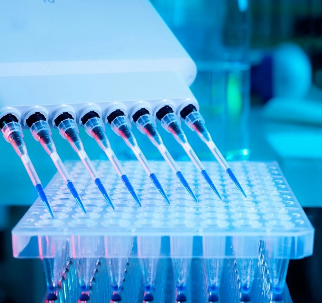
This assay is a quantitative sandwich ELISA. The immunoplate is pre- coated with a mouse monoclonal antibody specific for human PAI-1. Standards and samples are pipetted into the wells and any human PAI-1 present is bound by the immobilized antibody. After washing away any unbound substances, a biotin labelled polyclonal antibody specific for human PAI-1 is added to the wells. After wash step to remove any unbound reagents, streptavidin-HRP conjugate (STP-HRP) is added. After the last wash step, an HRP substrate solution is added and colour develops in proportion to the amount of human PAI-1 bound initially. The assay is stopped and the optical density of the wells determined using a microplate reader. Since the increases in absorbance are directly proportional to the amount of captured human PAI-1, the unknown sample concentration can be interpolated from a reference curve included in each assay.

